Widefield imaging for easier detection of peripheral pathologies
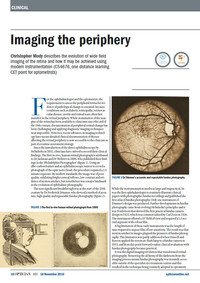
 The clinical utility of widefield fundus imaging is clear; it provides the eye care professional with the opportunity to support the diagnosis and monitor treatment and progression of diseases affecting the retinal periphery such as diabetic retinopathy, choroidal nevus, and cystoid macular edema.Today, the periphery can be visualized using a variety of imaging modalities including, MultiColor, autofluorescence, infrared reflectance and angiography, with simultaneous OCT.
This article explores the history of peripheral imaging and highlights how the latest widefield imaging technologies are providing clinicians with the opportunity to visualize more peripheral retinal structure than ever before.


