Three clinical cases in which ANTERION images make the difference
The high-resolution swept-source OCT images of ANTERION®* – the multimodal imaging platform for the anterior segment – provide visual confirmation and add reliability as well as accuracy in the diagnosis and follow-up of anterior segment alterations.
A picture is worth a thousand words. See for yourself …
 ANTERION and amniotic membrane grafts
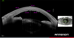
High-resolution images of the anterior segment provide the basis for accurate measurements. This image shows an amniotic membrane graft on an edematous and thick cornea. The ANTERION Metrics App allows you to calculate pre-defined anterior chamber parameters and perform free-hand measurements. In this case, the Metrics App assists you in determining the thickness of the amniotic membrane and of the patient’s cornea.
 ANTERION and femtosecond laser treatment
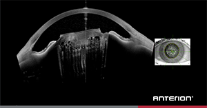
With the ANTERION imaging platform, you can visualize the entire anterior segment in great detail. This image was acquired after the use of femtosecond laser for cataract surgery and shows the laser capsulorhexis incision and nucleus fragmentation.
A NTERION and uveitis
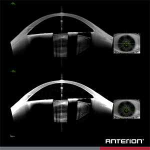
This patient with uveitis was imaged with ANTERION. In the OCT scans, you can see keratic precipitates, as hyperreflective dots on the posterior corneal surface. The iris atrophy allows for the imaging of the lens periphery.
By increasing the image contrast in the software, you can identify inflammation in the anterior chamber more easily.
Are you curious? Contact your local sales person for more info.
*ANTERION is not for sale in all countries.


