The retina up close with SPECTRALIS
Today we would like to share with you a journal article review and a recent study, showing how the SPECTRALIS® OCT Angiography Module (OCTA) and the High Magnification Module can support you to investigate the retina and aid in confident decision-making.

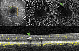
Microaneurysm in OCTA – Image Courtesy Dr. Rosa Dolz-Marco
Journal Article Review: "Microaneurysm visualization using five different optical coherence tomography angiography devices compared to fluorescein angiography"
The visualization of microaneurysms, which are considered the first detectable pathologic sign in patients with diabetic retinopathy, could play a key role in the detection of the disease. Parrulli et al. have investigated the usage of OCTA, providing detailed three-dimensional information of the different vascular layers, as a non-invasive alternative for the evaluation of microaneurysms.
"[…] OCTA could present a valuable alternative to FA when imaging microaneurysms. Within the scope of the current available devices used in this study, SPECTRALIS OCTA images detected a higher number of microaneurysms compared to the other OCTA devices."

Microstructures of the retina up close
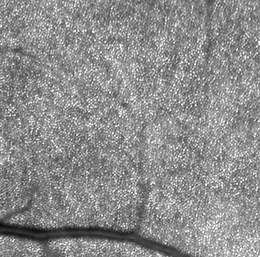
Study: "In vivo visualization of variable photoreceptor alteration in a case of peripapillary congenital hypertrophy of the retinal pigment epithelium using SPECTRALIS high magnification module"
A recent study by Elahi et al. shows the potential of the SPECTRALIS High Magnification Module for visualizing variable photoreceptor alteration in peripapillary congenital hypertrophy.
It highlights that an in vivo visualization of photoreceptors "[…] would represent an important breakthrough in retinal imaging, and perhaps lead to a better understanding of retinal diseases’ pathophysiology."
If you are interested in finding out more about potential applications for the different modules of the Heidelberg Engineering SPECTRALIS please visit the Heidelberg Engineering Business Lounge.


