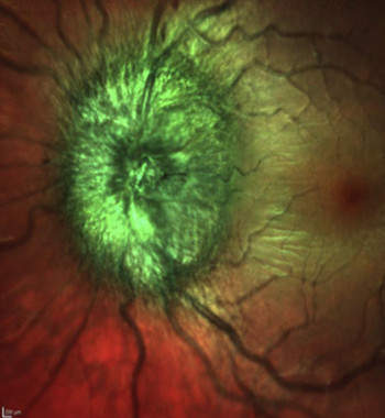
Journal Article Review – Use of MultiColor imaging in the assessment of suspected papilledema in 20 consecutive children
 True papilledema or pseudopapilledema?
Differentiating between these entities in children based on noninvasive imaging would be greatly preferable over an invasive neurological examination – the current gold standard. The authors of a recent study investigated whether the SPECTRALIS® MultiColor Module is helpful to make this distinction. Taking MultiColor images from the optic nerve head of 20 consecutive patients delivered very promising results.
The SPECTRALIS MultiColor Module uses three laser wavelengths simultaneously to provide diagnostic images that show distinct structures at different depths within the retina. The high-resolution, detailed MultiColor images can highlight structures and pathologies not visible on ophthalmoscopy and fundus photography. MultiColor images may even be acquired in patients with cataracts or nystagmus. Available only on the SPECTRALIS platform, the combination of MultiColor with OCT brings a new dimension of detail and versatility to ophthalmic imaging.
Please find other Journal Article Reviews here.*
*Business Lounge login required.
Please note that the SPECTRALIS is not FDA cleared for clinical use in a pediatric population in the USA.


