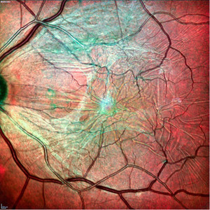
Article Review – Visualization of macular pucker by MultiColor scanning laser imaging

by Muftuoglu IK, Bartsch DU, Barteselli G, Gaber R, Nezgoda J, Freeman WR
A multimodal imaging approach can empower clinicians to detect and differentiate between retinal diseases. The SPECTRALIS® MultiColor Module captures a composite of three monochromatic reflectance images to deliver high-resolution detail of retinal structure at different depths.
A recent study quantitatively assessed the value of MultiColor compared to color fundus photography (CFP) and OCT in detecting and visualizing macular epiretinal membranes (ERM).
FIND OUT MORE ABOUT THE RESULTS
As part of a multimodal approach, the SPECTRALIS MultiColor Module can be combined with other imaging modalities to support the clinician’s diagnostic confidence. Using three laser wavelengths simultaneously, the MultiColor Module provides diagnostic images that show distinct structures at different depths within the retina. The high-resolution SPECTRALIS MultiColor images can highlight structures and pathologies not visible on ophthalmoscopy and fundus photography. The images may even be acquired in patients with cataracts or nystagmus. The combination of MultiColor with OCT brings together greater detail and more information in a single platform.


